|
Miltenyi Biotec
cd43 Cd43, supplied by Miltenyi Biotec, used in various techniques. Bioz Stars score: 94/100, based on 1 PubMed citations. ZERO BIAS - scores, article reviews, protocol conditions and more https://www.bioz.com/product/anti+cd43/CD43+Antibody%2C+anti-rat%2C+REAfinity/10__17816_slash_morph__681777-98-28-30 Average 94 stars, based on 1 article reviews
cd43 - by Bioz Stars,
2026-09
94/100 stars
|
Buy from Supplier |
|
Miltenyi Biotec
cd43 pe 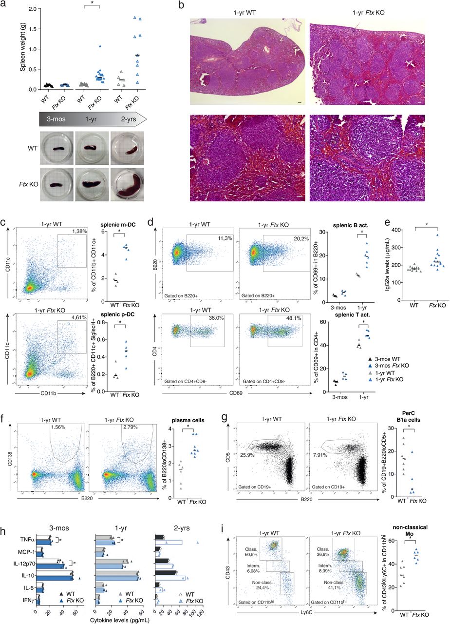 Cd43 Pe, supplied by Miltenyi Biotec, used in various techniques. Bioz Stars score: 92/100, based on 1 PubMed citations. ZERO BIAS - scores, article reviews, protocol conditions and more https://www.bioz.com/product/anti+cd43/CD43+Antibody%2C+anti-mouse%2C+REAfinity/bio_rxiv__2023__04__20__537662-150-57-59 Average 92 stars, based on 1 article reviews
cd43 pe - by Bioz Stars,
2026-09
92/100 stars
|
Buy from Supplier |
|
Bio-Rad
w3 13 W3 13, supplied by Bio-Rad, used in various techniques. Bioz Stars score: 93/100, based on 1 PubMed citations. ZERO BIAS - scores, article reviews, protocol conditions and more https://www.bioz.com/product/anti+cd43/Mouse+anti+Rat+CD43/pmc01809229-118-16-20 Average 93 stars, based on 1 article reviews
w3 13 - by Bioz Stars,
2026-09
93/100 stars
|
Buy from Supplier |
|
Miltenyi Biotec
anti cd43 antibody  Anti Cd43 Antibody, supplied by Miltenyi Biotec, used in various techniques. Bioz Stars score: 90/100, based on 1 PubMed citations. ZERO BIAS - scores, article reviews, protocol conditions and more https://www.bioz.com/product/anti+cd43/CD43+Antibody%2C+anti-mouse/pmc09401627-196-11-13 Average 90 stars, based on 1 article reviews
anti cd43 antibody - by Bioz Stars,
2026-09
90/100 stars
|
Buy from Supplier |
|
fluidigm
anti mouse cd43 clone s11 146nd  Anti Mouse Cd43 Clone S11 146nd, supplied by fluidigm, used in various techniques. Bioz Stars score: 90/100, based on 1 PubMed citations. ZERO BIAS - scores, article reviews, protocol conditions and more https://www.bioz.com/product/anti+cd43/Anti-Mouse+CD43+(S11)-146Nd/pmc06591003-4-0-5 Average 90 stars, based on 1 article reviews
anti mouse cd43 clone s11 146nd - by Bioz Stars,
2026-09
90/100 stars
|
Buy from Supplier |
|
Miltenyi Biotec
anti cd23 microbeads Anti Cd23 Microbeads, supplied by Miltenyi Biotec, used in various techniques. Bioz Stars score: 97/100, based on 1 PubMed citations. ZERO BIAS - scores, article reviews, protocol conditions and more https://www.bioz.com/product/anti+cd43/CD43+Glycoform+Antibody%2C+anti-mouse%2C+REAfinity/pm37095377-558-15-18 Average 97 stars, based on 1 article reviews
anti cd23 microbeads - by Bioz Stars,
2026-09
97/100 stars
|
Buy from Supplier |
|
Miltenyi Biotec
cd43 apc miltenyi biotec df t1 Cd43 Apc Miltenyi Biotec Df T1, supplied by Miltenyi Biotec, used in various techniques. Bioz Stars score: 90/100, based on 1 PubMed citations. ZERO BIAS - scores, article reviews, protocol conditions and more https://www.bioz.com/product/anti+cd43/CD43+Antibody%2C+anti-human/pmc05940870__41467_2018_4134_MOESM1_ESM-51-154-156 Average 90 stars, based on 1 article reviews
cd43 apc miltenyi biotec df t1 - by Bioz Stars,
2026-09
90/100 stars
|
Buy from Supplier |
|
Bio-Rad
mca1096ga  Mca1096ga, supplied by Bio-Rad, used in various techniques. Bioz Stars score: 90/100, based on 1 PubMed citations. ZERO BIAS - scores, article reviews, protocol conditions and more https://www.bioz.com/product/anti+cd43/Mouse+anti+Sheep+CD43/pmc09728927-11-9-7 Average 90 stars, based on 1 article reviews
mca1096ga - by Bioz Stars,
2026-09
90/100 stars
|
Buy from Supplier |
|
Bio-Rad
rat anti mouse cd43 Rat Anti Mouse Cd43, supplied by Bio-Rad, used in various techniques. Bioz Stars score: 93/100, based on 1 PubMed citations. ZERO BIAS - scores, article reviews, protocol conditions and more https://www.bioz.com/product/anti+cd43/Rat+anti+Mouse+CD43/pm18714009-70-109-132 Average 93 stars, based on 1 article reviews
rat anti mouse cd43 - by Bioz Stars,
2026-09
93/100 stars
|
Buy from Supplier |
|
fluidigm
3150006b  3150006b, supplied by fluidigm, used in various techniques. Bioz Stars score: 96/100, based on 1 PubMed citations. ZERO BIAS - scores, article reviews, protocol conditions and more https://www.bioz.com/product/anti+cd43/Anti-Human+CD43+(84-3C1)-150Nd/pmc08776501-133-6-3 Average 96 stars, based on 1 article reviews
3150006b - by Bioz Stars,
2026-09
96/100 stars
|
Buy from Supplier |
|
Miltenyi Biotec
miltenyi biotech 130 114 749 cd47 fitc Miltenyi Biotech 130 114 749 Cd47 Fitc, supplied by Miltenyi Biotec, used in various techniques. Bioz Stars score: 93/100, based on 1 PubMed citations. ZERO BIAS - scores, article reviews, protocol conditions and more https://www.bioz.com/product/anti+cd43/CD43+Antibody%2C+anti-human%2C+REAfinity/pmc06764950__41467_2019_12408_MOESM1_ESM-88-131-131 Average 93 stars, based on 1 article reviews
miltenyi biotech 130 114 749 cd47 fitc - by Bioz Stars,
2026-09
93/100 stars
|
Buy from Supplier |
|
Bio-Rad
mouse anti human cd43  Mouse Anti Human Cd43, supplied by Bio-Rad, used in various techniques. Bioz Stars score: 88/100, based on 1 PubMed citations. ZERO BIAS - scores, article reviews, protocol conditions and more https://www.bioz.com/product/anti+cd43/Mouse+anti+Human+CD43/pm19556303-29-16-21 Average 88 stars, based on 1 article reviews
mouse anti human cd43 - by Bioz Stars,
2026-09
88/100 stars
|
Buy from Supplier |
Image Search Results
Journal: bioRxiv
Article Title: Altered X-chromosome inactivation predisposes to autoimmune manifestations in mice
doi: 10.1101/2023.04.20.537662
Figure Lengend Snippet: a. Spleen weight of wild-type (WT) and Ftx KO females at 3-months, 1-year and 2-years of age. Median values are shown. ( t-test , * p -values < 0.05). Underneath, representative images of WT and Ftx KO spleens from 3-month, 1-year and 2-year-old females. b. Representative images of hematoxylin-eosin staining on sections of spleens from 1-year-old WT and Ftx KO females. Scale bar; 100 μm. c. Representative flow cytometry analysis of splenic myeloid dendritic cells (m-DC) in WT and Ftx KO 1-year-old females. On the right, percentages of splenic m-DC (CD11b + CD11c + ) and splenic plasmacytoid dendritic cells (p-DC) (CD11c + B220 + SiglecH + ) in leucocytes. Each triangle represents a mouse. Median values are shown. ( t-test , * p -values < 0.05). d. Representative flow cytometry analysis of spontaneously activated B cells (B220 + CD69 + ) (upper panels) or of spontaneously activated T cells (CD4 + CD69 + ) (lower panels) in spleen from 1-year-old WT and Ftx KO females. Percentages in leucocytes are shown on the graphs beneath. Each triangle represents a mouse. Median values are shown. ( t-test , * p -values < 0.05). e. IgG2a natural antibody levels in sera of 1-year- and 2-year-old WT or Ftx KO females measured by ELISA. Each triangle represents a mouse. Median values are shown. ( t-test , * p -values < 0.05). f. Representative flow cytometry analysis of (B220 + CD138 + ) plasma cells in the spleen of 1-year-old WT and Ftx KO females. Percentages in leucocytes are shown on the graphs beneath. Each triangle represents a mouse. Median values are shown. ( t-test , * p -values < 0.05). g. Representative flow cytometry analysis of (CD19 + B220 lo CD5 + ) natural antibody producing B1a in the peritoneal cavity (PerC) of 1-year-old WT and Ftx KO females. Percentages in leucocytes are shown on the graphs beneath. Each triangle represents a mouse. Median values are shown. ( t-test , * p -values < 0.05). h. Cytokines levels in the blood analysed with CBA assays on sera from 3-month-, 1-year-, or 2-year-old WT and Ftx KO females. Each triangle represents a mouse. Median values are shown. ( t-test , * p -values < 0.05). i. Representative flow cytometry analysis of monocyte populations including non-classical (CD11b hi CD43 lo Ly6C + ) scavenger monocytes in the spleen of 1-year-old WT and Ftx KO females. Percentages in leucocytes are shown on the graphs beneath. Each triangle represents a mouse. Median values are shown. ( t-test , * p -values < 0.05).
Article Snippet: Bone marrow, spleen, blood and peritoneal cavity cells were stained using the following antibodies: CD3 PerCP-Vio770 (130-119-656, Miltenyi Biotec), CD4-APC (130-123-207, Miltenyi Biotec), CD5-APC-Vio770 (130-120-165, Miltenyi Biotec), CD8-FITC (130-118-468, Miltenyi Biotec), CD11b APC (553312, BD Pharmingen), CD11c PE-Vio770 (130-110-840, Miltenyi Biotec), CD19-FITC (557398, BD Pharmingen), CD21-APC-Vio770 (130-111-733, Miltenyi Biotec), CD23-PE-Vio770 (130-118-764, Miltenyi Biotec), CD38-PE (130-123-571, Miltenyi Biotec),
Techniques: Staining, Flow Cytometry, Enzyme-linked Immunosorbent Assay, Clinical Proteomics
Journal: Science Advances
Article Title: AFF3, a susceptibility factor for autoimmune diseases, is a molecular facilitator of immunoglobulin class switch recombination
doi: 10.1126/sciadv.abq0008
Figure Lengend Snippet: ( A ) Serum antibodies from Aff3 flox/flox Cd4cre and Cd19cre mice were measured by ELISA ( n = 5 in each group). ( B ) Flow cytometry analysis of class switching to IgG1. CD43 − B cells were harvested from Aff3 +/− (purple) or Aff3 −/− (red) mice and cultured with LPS and IL-4 (10 μg/ml). After 4 days, the cells were analyzed by flow cytometry. The panels show the IgG1 and IgG3 expression profiles and the statistical analysis ( n = 4 in each group). ( C ) Flow cytometry analysis of class switching to IgG2c. CD43 − B cells were cultured with LPS and IFN-γ (10 μg/ml). The panels show the IgG2c expression profiles and the statistical analysis ( n = 3 in each group). ( D ) Overexpression of the AFF3 compensated for the Aff3 −/− phenotype. CD43 − B cells were harvested from Aff3 −/− mice and cultured with LPS and IL-4. The cDNAs of Aff3 , Aff1 , and Aff4 were transduced with a retroviral vector carrying the green fluorescent protein (GFP) marker. The panel shows the percentage of IgG1 + cells in GFP + CD19 + cells ( n = 3 in each group). EV, empty vector. The data are shown as means ± SEM. * P < 0.05, *** P < 0.005, and **** P < 0.001. The P values were calculated using unpaired t test with Welch’s correction in (A to C) and via one-way ANOVA with Dunnett’s multiple comparisons test in (D). The data shown in this figure are representative of at least two experiments.
Article Snippet: Naïve B cells were purified by negative selection using a biotin-conjugated
Techniques: Enzyme-linked Immunosorbent Assay, Flow Cytometry, Cell Culture, Expressing, Over Expression, Transduction, Retroviral, Plasmid Preparation, Marker

Journal: Science Advances
Article Title: AFF3, a susceptibility factor for autoimmune diseases, is a molecular facilitator of immunoglobulin class switch recombination
doi: 10.1126/sciadv.abq0008
Figure Lengend Snippet: ( A ) CD43 − B cells were stained with CFSE and cultured. The cells were analyzed by flow cytometry. ( B ) Transcriptome analysis of Aff3 -deficient B cells. CD43 − B cells were cultured with LPS and IL-4. After 3 days of culture, total RNA was prepared and analyzed on a cDNA microarray. ( C ) Quantitative reverse transcription polymerase chain reaction (qRT-PCR) and Western blotting analyses of Aicda ( n = 3 in each group for RT-PCR). CD43 − B cells were cultured with LPS (10 μg/ml) and IL-4 (10 ng/ml). Western blotting was performed with anti-AID antibody, and the membrane was reprobed with anti–extracellular signal–regulated kinase 1/2 (ERK1/2) antibody to assess the loading amounts. nt, nucleotide. ( D ) AFF3 binds to switch regions. CD43 − B cells were cultured with LPS (10 μg/ml) and IL-4 (10 ng/ml) for 3 days. Empty (purple) or 3xFLAG-tagged cDNA of the C-terminal half of Aff3 (red) was transduced. The efficiency of transduction was estimated on the basis of the expression of enhanced GFP (EGFP), which was coexpressed with AFF3 by IRES (internal ribosomal entry site) insertion. Then, the cells were analyzed by ChIP with an anti-FLAG antibody. The amount of DNA precipitated was measured by real-time PCR ( n = 3 in each group), with primers specific to the switch regions or the Acta (encoding α-actin) gene serving as a negative control. Sμ-SR indicates the switch region of IgM (table S4) . Sγ1-c and Sγ1-d indicate the switch region of IgG1 (fig. S7 and table S4). In (C and D), the data are shown as means ± SEM. **** P < 0.001. The P values were calculated using unpaired t tests with Welch’s correction. The data shown in this figure are representative of at least two experiments except for those in (B). The primers used in the experiments are listed in table S4.
Article Snippet: Naïve B cells were purified by negative selection using a biotin-conjugated
Techniques: Staining, Cell Culture, Flow Cytometry, Microarray, Reverse Transcription, Polymerase Chain Reaction, Quantitative RT-PCR, Western Blot, Reverse Transcription Polymerase Chain Reaction, Membrane, Transduction, Expressing, Real-time Polymerase Chain Reaction, Negative Control

Journal: Science Advances
Article Title: AFF3, a susceptibility factor for autoimmune diseases, is a molecular facilitator of immunoglobulin class switch recombination
doi: 10.1126/sciadv.abq0008
Figure Lengend Snippet: ( A ) qRT-PCR was used to analyze the germline and postswitch transcript levels of IgG1 ( n = 3 in each group). Aff3 +/− (purple) or Aff3 −/− (red) CD43 − B cells were cultured with LPS (10 μg/ml) and the indicated amount of IL-4. After 3 days of culture, total RNA was extracted from the cells and analyzed. ( B ) Semiquantitative PCR was performed to analyze CSR. CD43 B cells were cultured as described in (A), and genomic DNA was extracted. PCR of the IgM and IgG1 switch regions in combination was performed with the indicated primers. The genomic DNA was serially diluted and used as the template for PCR. Primers for CD19 were used as an internal control. ( C ) Genomic DNA was prepared as described in (B). Fragments of IgM-IgG1 chimeric switch regions were amplified by nested PCR. The fragments were sequenced, and the mutation rates per nucleotide were plotted ( n = 3 in each group). The detailed data are shown in table S1. In (A and C), the data are shown as means ± SEM. * P < 0.05 and *** P < 0.005. The P values were calculated using unpaired t tests with Welch’s correction. The data in (A and B) are representative of at least two experiments. The primers used are listed in table S4.
Article Snippet: Naïve B cells were purified by negative selection using a biotin-conjugated
Techniques: Quantitative RT-PCR, Cell Culture, Control, Amplification, Nested PCR, Mutagenesis
Journal: Immunity
Article Title: Pro-inflammatory Aorta-Associated Macrophages Are Involved in Embryonic Development of Hematopoietic Stem Cells
doi: 10.1016/j.immuni.2019.05.003
Figure Lengend Snippet:
Article Snippet:
Techniques: Purification, Recombinant, Software, Control, High Throughput Screening Assay, Sequencing
Journal: PLOS ONE
Article Title: B cell phenotypes and maturation states in cows naturally infected with Mycobacterium avium subsp. Paratuberculosis
doi: 10.1371/journal.pone.0278313
Figure Lengend Snippet: Primary and secondary antibodies used to discriminate B cell subpopulations.
Article Snippet: CD43 - , , CO.44B8-IgG 1 ,
Techniques: Marker
Journal: Cell
Article Title: A blood atlas of COVID-19 defines hallmarks of disease severity and specificity
doi: 10.1016/j.cell.2022.01.012
Figure Lengend Snippet:
Article Snippet: CD43 (84-3C1)-150Nd ,
Techniques: Mass Cytometry, Flow Cytometry, Recombinant, Staining, Selection, Antibody Labeling, Labeling, Isolation, Sample Prep, Luminex, Quantitative Proteomics, Generated, Gene Expression, Clone Assay, Marker, Expressing, Mass Spectrometry, Derivative Assay, RNA Sequencing, Sequencing, Illumina Sequencing, Software, Variant Assay
Journal: International immunology
Article Title: Anti-moesin antibodies derived from patients with aplastic anemia stimulate monocytic cells to secrete TNF-alpha through an ERK1/2-dependent pathway.
doi: 10.1093/intimm/dxp058
Figure Lengend Snippet: Fig. 1. The specific induction of TNF-a from THP-1 cells and their phenotypic changes by anti-moesin pAb treatment. (A) Left panel: dose- dependent stimulation of THP-1 cells by anti-moesin pAbs. THP-1 cells were cultured for 48 h in the presence of several concentrations of anti- moesin pAbs or human IgG from healthy individuals or anti-human CD43 mAb. TNF-a concentration in the culture supernatant was measured by ELISA. *P < 0.01, **P < 0.001. The figure shows the representative results of three independent experiments. Right panel: THP-1 cells were stained with FITC-labeled anti-moesin mAb (upper panel, filled histogram), anti-CD43 mAb (bottom panel, filled histogram) or isotype antibodies (open histogram). (B) The effect of anti-moesin pAb treatment on the surface marker expression on THP-1 cells. THP-1 cells were cultured for 48 h in the presence or absence of 5 lg ml1 of anti-moesin pAbs or isotype human IgG, and the expression levels of several cell surface proteins were determined by flow cytometry. Filled histogram, untreated cells; green, cells stained with isotype control antibodies; red, human IgG-stimulated cells; blue, anti-moesin pAb-stimulated cells. The figure shows the representative results of three independent experiments.
Article Snippet: Mouse anti-human Toll-like receptor 4 (TLR4) antibody was purchased from Abcam Inc. (Cambridge, MA, USA) and
Techniques: Cell Culture, Concentration Assay, Enzyme-linked Immunosorbent Assay, Staining, Labeling, Marker, Expressing, Cytometry, Control